In other words, you need a system!
In the end, whatever works for you is important, but this is one I learned: Technical ABCDE.
Technical
This is all the non-anatomy parts of the film.
ID: name, type of image, position of image (AP vs PA), correct date and MRN tend to be skipped, though some attendings will like to hear them. You should still verify these regardless.
Image: Inspiration (can you count 8-10 posterior ribs), penetration (usually less of an issue with digitally captured x-rays), and rotation (look for symmetric angle of clavicles with sternum in non-rotated film).
Patient: Any tubes, wires, catheters you see - especially note where central lines, PICC lines, and ET tubes end.
A
Airway: Is the trachea midline or deviated?
B
Bones: Look for overt fractures, dislocations, and lytic lesions
C
Cardiac: Check heart size (less than half the width of the chest space is normal - AP films are not as reliable as PA films for this), silhouette and edges, mediastinum and aortic knob.
D
Diaphragm: Check for a right hemidiaphragm (right diaphragm elevated), sharp costophrenic angles, and air under the diaphragm (including normal gastric bubble).
E
Everything else (soft tissues): Look for soft tissue swelling or mass.
After you've done that, then and only then do you proceed to the paydirt: the lung fields. Check for lung marking to the perimeter, opacities, masses, haziness. Especially in EM don't forget to check the apices for pneumothorax.
The idea is that you delay looking at the thing you are naturally drawn to, the reason you got the CXR, until you've taken care of everything else. How can you have any pudding if you don't eat your meat?
Further reading:
This isn't something you learn from one blog post. It takes repetition and actually reading x-rays. The green chart below will link to a LITFL post on the same basics with a slightly different mnemonic. A video lecture with images can be found here.


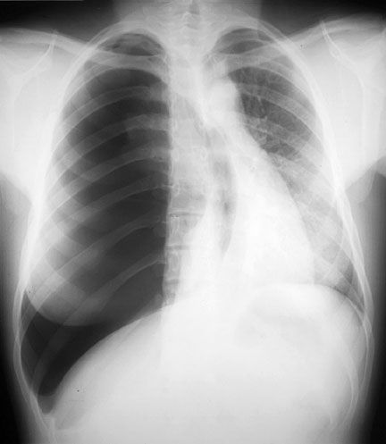
Double secret bonus: You never want to take the leading CXR in this post - it is a tension pneumothorax, which should have been diagnosed clinically and treated immediately since it is an emergency. Don't delay for an x-ray!
Precise and to the point, thanks for sharing such informative blog with us. Looking forward to see more such informative posts from you
ReplyDelete